Chapter 5. The Region Lens
Johnson
2026-08-15
The coarsest tier of the ladder is also, in some ways, the most instructive: GeoMx. The 48 GeoMx samples are region-of-interest profiles, each one a transcriptomic snapshot of a user-selected tissue region, measured at ~17,000–19,000 probes. There are no coordinates. There is no spatial graph. There is, in many analyses, an assumption that such data are barely “spatial” at all.
This chapter is about what region-level biology can still say. The cohort is a coherent one: all 48 ROIs come from a single kidney-transplant study, and each ROI carries a compartment label (glomerulus, peritubular capillary, or vessel). I do not use those labels to analyze; I use them afterward, to check.
Scoring a region for nephron programs
The approach is the same module scoring used on single cells as mentioned earlier, applied to a region: each ROI is scored for the eighteen nephron programs. A region is a mixture, so the scores are composition proxies, but as this table shows, they are informative ones.
The scoring is the same gene-set step used throughout this analysis, applied here to a region transcriptome rather than to cells, and the compartment labels that came with the cohort were held aside during scoring and consulted only afterward. That ordering is what makes the agreement in the table a real result rather than a circular one.
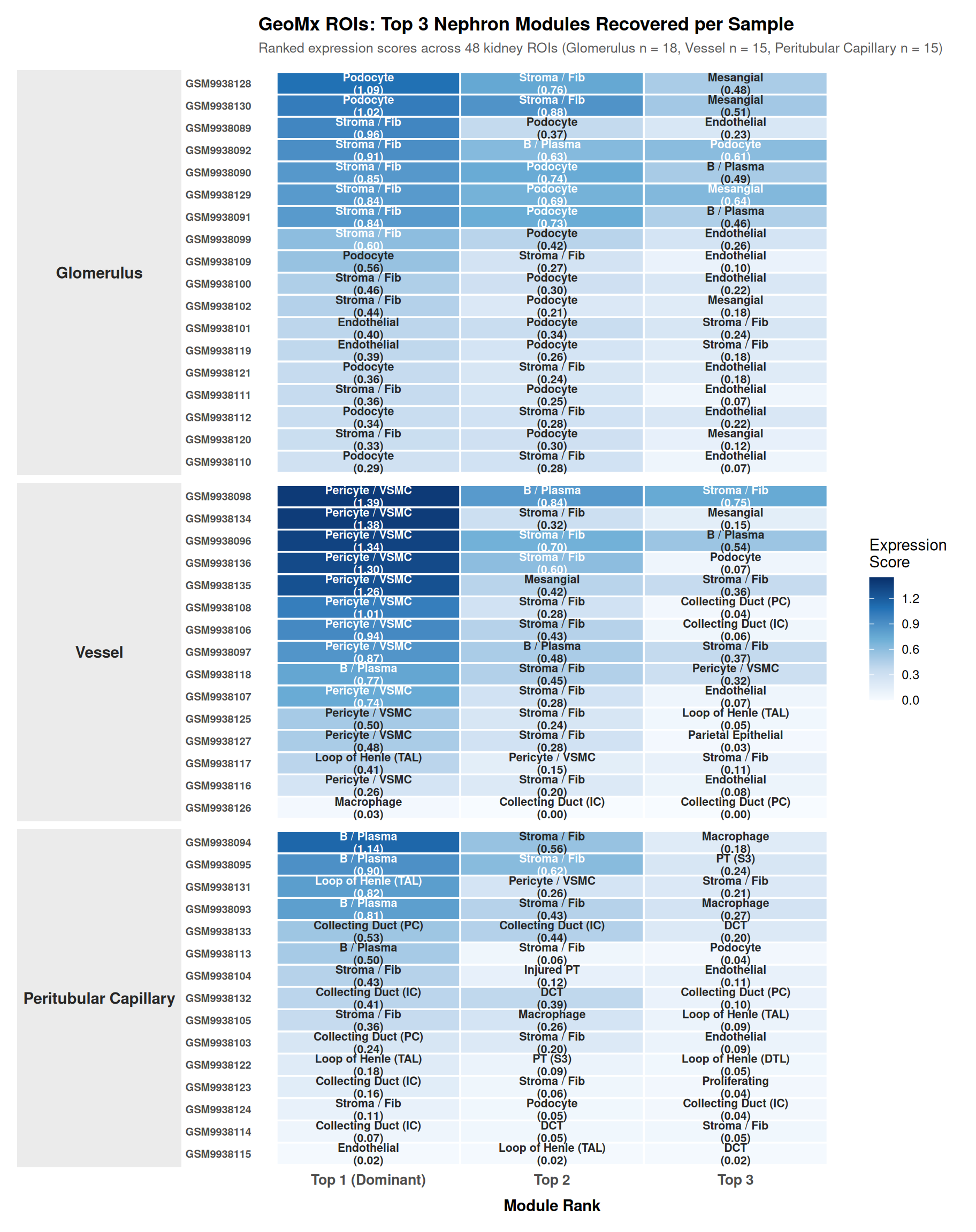
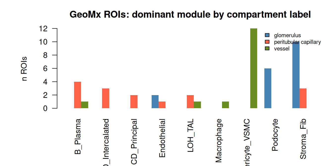
The pattern across all 48 ROIs:
Vessel ROIs are pericyte/vascular-smooth-muscle-dominant (12 of 14). The vessel program (ACTA2, RGS5, MYH11) is the signature of the vascular wall.
Glomerular ROIs are podocyte- and stroma-enriched.
Peritubular-capillary ROIs carry immune and intercalated-cell signatures.
This is compartment identity, recovered from expression alone, with no coordinates. A region transcriptome reflects the identity of the tissue from which it was collected.
Why this matters
The region lens is the one with the clearest path to the clinic. GeoMx is fast, comparatively cheap, and clinically scalable; a hospital system can profile hundreds of annotated regions from archived tissue. The standard objection is that without coordinates it is “not really spatial.” This chapter’s answer is that the biology of a region is still in its transcriptome: a vessel ROI’s pericyte dominance, a glomerular ROI’s podocyte program, and a capillary ROI’s immune signature are all recoverable with no spatial information beyond the user’s own selection.
In a transplant-rejection context, which is what this cohort is, that matters directly. Chronic antibody-mediated rejection attacks the peritubular capillary bed; the compartment architecture that the modules reconstruct is precisely the architecture under attack. The region lens cannot indicate where within a region the biology changes, but it can indicate, at scale, which compartments carry which disease signatures. That is a real analytical capability, and it is the region rung’s contribution to this analysis.
The limits of the region lens
A region is a mixture. The module scores are composition proxies, not cell identities; they cannot resolve a podocyte from a parietal cell within a glomerular ROI.
No spatial graph, no neighborhoods, no domains. Every spatial analysis in this analysis that needs coordinates simply does not apply to GeoMx, and I do not pretend otherwise.
One ROI per sample. There is no within-sample spatial structure at all; each sample is a single point.
What the region lens establishes, then, is a clean and useful fact: compartment identity survives region-level profiling, with no coordinates, at clinical scale. The rest of this analysis treats GeoMx accordingly, as a coarser, larger-scale companion to the single-cell and spot lenses, able to answer compartment-level questions across many samples.